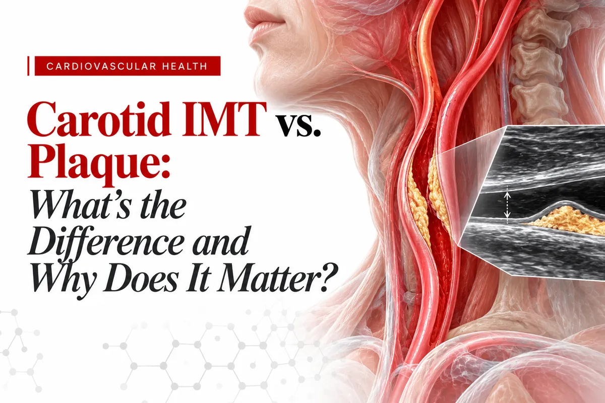
Carotid IMT vs. Plaque: What's the Difference and Why Does It Matter?
Vasolabs | Carotid IMT vs. Plaque
Understanding what your carotid ultrasound reveals about your artery walls and cardiovascular health.
When you receive a medical report, understanding the numbers and terminology can sometimes feel overwhelming. Terms such as carotid intima-media thickness (IMT) and carotid plaque may appear together, but they describe two different findings.
Both provide information about the condition of your arteries, yet they measure different aspects of vascular health.
Carotid IMT measures the thickness of the artery wall, while carotid plaque identifies localized areas of buildup within the artery.
Understanding this distinction can help you ask better questions, have more informed conversations with your healthcare provider, and take a more active role in your cardiovascular health.
Let's explore what each measurement means, why the distinction matters, and how carotid ultrasound findings may contribute to a broader cardiovascular prevention strategy.
Understanding Your Carotid Arteries
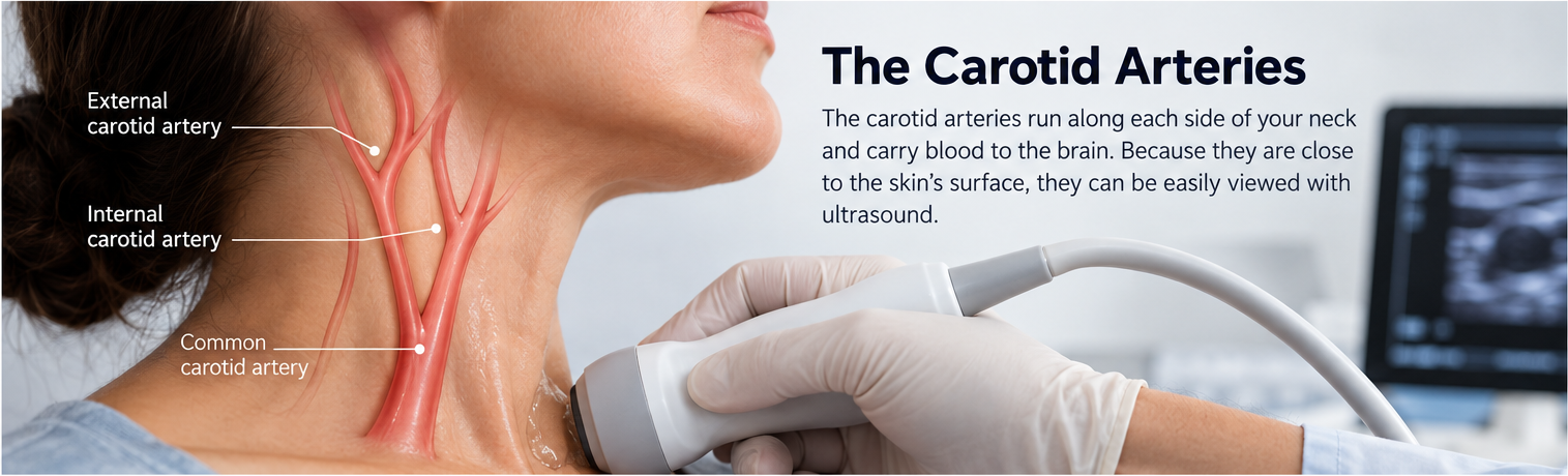
Your carotid arteries are two major blood vessels located on either side of your neck. Their primary function is to deliver oxygen rich blood to your brain, head, and face.
Like other arteries throughout your body, the carotid arteries have walls made of several layers that help maintain blood flow and support healthy circulation.
These layers include:
Intima: The innermost layer, which provides a smooth surface for blood to flow through.
Media: The middle layer, composed primarily of smooth muscle that helps regulate blood vessel function.
Adventitia: The outer layer, which provides structural support and protection.
Over time, these arteries may undergo changes associated with aging, blood pressure, cholesterol levels, smoking, diabetes, and other cardiovascular risk factors.
Some changes involve gradual thickening of the artery wall. Others involve the development of atherosclerotic plaque.
Because the carotid arteries are relatively close to the skin's surface, healthcare professionals can examine them using ultrasound technology.
Carotid ultrasound is a noninvasive imaging technique that uses sound waves to visualize the arteries without radiation, needles, or contrast dye.
Depending on the type of examination performed, it can provide information about artery wall thickness, plaque, and blood flow.
What Is Carotid Intima-Media Thickness (IMT)?
Carotid Intima-Media Thickness, commonly abbreviated as CIMT or IMT, refers to the combined thickness of the intima and media layers of the carotid artery wall.
During an ultrasound examination, these layers can be visualized and measured in millimeters.
Researchers have studied CIMT for decades to better understand changes in artery structure and their relationship to cardiovascular risk.
What Can Influence IMT?
Several factors may affect carotid artery wall thickness, including:
Age: Artery walls tend to become thicker as people get older.
High blood pressure: Increased pressure places additional mechanical stress on artery walls.
Cholesterol and metabolic health: Abnormal lipid levels and metabolic conditions may contribute to vascular changes.
Diabetes: Elevated blood sugar and related metabolic changes can affect blood vessel health.
Smoking: Tobacco exposure can damage blood vessels and contribute to cardiovascular disease.
Family history: Genetic factors can influence cardiovascular risk and vascular characteristics.
These factors do not affect everyone in exactly the same way.
A higher IMT measurement does not automatically mean that a person has significant plaque buildup or blocked arteries.
Instead, it provides information about artery wall structure that must be interpreted alongside other health factors.
What Does an IMT Number Mean?
IMT measurements are typically expressed in millimeters.
When appropriate reference data are available, a clinician may compare an individual's measurement with values observed in people of similar age and sex, sometimes accounting for additional population characteristics.
For example, an IMT measurement that is expected in an older adult may have a different interpretation in a younger individual.
However, there is no single IMT number that can reliably determine cardiovascular health for everyone.
Measurement technique, the artery segment examined, reference ranges, and the person's overall medical history all influence interpretation.
Importantly, current cardiovascular prevention guidelines do not recommend routine CIMT measurement as a standalone test for predicting a first cardiovascular event. Its value depends on the clinical context.
What Is Carotid Plaque?
While IMT describes the thickness of the artery wall, carotid plaque refers to a more localized structural change.
Carotid plaque is a focal buildup within the artery wall associated with atherosclerosis.
Atherosclerosis is a process in which cholesterol containing particles, inflammatory cells, connective tissue, calcium, and other materials accumulate within the arterial wall.
As plaque develops, it may change the shape or thickness of a specific area of the artery.
Unlike a general IMT measurement, plaque represents a distinct area of atherosclerotic change.
What Can a Carotid Ultrasound Reveal About Plaque?
Depending on the imaging protocol and equipment used, a carotid ultrasound may help identify:
Plaque presence: Whether visible plaque has developed.
Plaque location: Where plaque is located within the carotid arteries.
Plaque size: The measured dimensions of a visible plaque.
Plaque appearance: Certain ultrasound characteristics, such as areas that appear darker, brighter, or calcified.
Artery narrowing: Whether plaque is associated with narrowing of the vessel, particularly when a carotid duplex study is performed.
Not all plaques have the same appearance or characteristics.
Some may contain more calcium, while others may contain more lipid rich or softer tissue components.
Ultrasound can describe certain visible characteristics, but it cannot determine every detail of plaque composition or reliably predict whether a particular plaque will rupture.
Does Finding Plaque Mean Something Is Wrong?
The presence of carotid plaque is evidence of atherosclerosis and may be associated with increased cardiovascular risk.
However, finding plaque does not automatically mean that a heart attack or stroke is about to happen.
Many individuals with carotid plaque have no symptoms.
The significance of a finding depends on factors such as plaque burden, degree of narrowing, symptoms, medical history, and overall cardiovascular risk.
Your healthcare provider can help determine whether a plaque finding warrants further evaluation or changes to your prevention plan.
Carotid IMT vs. Plaque: Understanding the Key Differences
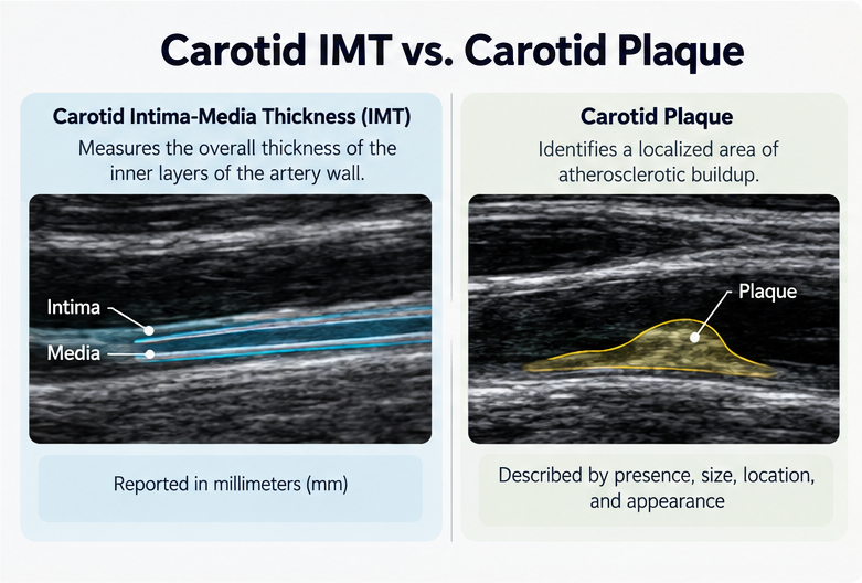
Although both findings involve the artery wall, they answer different questions.
Carotid IMT | Carotid Plaque |
Measures the combined thickness of the intima and media layers | Identifies a localized area of atherosclerotic buildup |
Usually expressed in millimeters | May be described by presence, size, location, and appearance |
Can increase with aging and other vascular influences | Indicates a focal atherosclerotic change |
Does not necessarily indicate plaque | Can be present even when IMT is within an expected range |
Has limited value for routine cardiovascular risk prediction | Can provide additional information for cardiovascular risk assessment |
The simplest way to remember the difference is this: IMT measures artery wall thickness, while plaque identifies specific areas of buildup.
A person can have an IMT measurement within an expected range and still have carotid plaque.
Likewise, someone may have increased artery wall thickness without a distinct plaque being identified.
This is why IMT and plaque should not be treated as interchangeable findings.
Can You Have Normal IMT but Still Have Plaque?
Yes!
One of the important distinctions between IMT and plaque is that they do not necessarily develop or appear in the same way.
An individual may have an IMT measurement that falls within an expected reference range while still having a localized plaque elsewhere in the carotid artery.
This happens because IMT is typically measured in a standardized region of the artery wall, whereas plaque can develop in other locations, including areas where the artery branches.
The opposite is also possible.
An individual may have an increased IMT measurement without visible focal plaque.
In these cases, artery wall thickening may be influenced by aging, blood pressure, or other vascular changes rather than a clearly identifiable plaque deposit.
A normal IMT measurement does not necessarily mean that the carotid arteries are free of plaque.
Evaluating both findings may provide a more detailed description of carotid artery structure, although the additional clinical value depends on the individual and the reason for testing.
Why Does the Difference Matter for Cardiovascular Prevention?
Cardiovascular disease can develop gradually, sometimes without noticeable symptoms.
Traditional risk assessments consider factors such as:
Blood pressure
Cholesterol levels
Blood sugar and diabetes status
Smoking history
Age
Family history
Existing medical conditions
These factors help healthcare professionals estimate a person's likelihood of developing cardiovascular disease.
Imaging can sometimes provide additional information by showing structural changes that have already occurred in the arteries.
For example, identifying carotid plaque may help a clinician better understand a person's atherosclerotic disease burden, especially when a prevention decision is uncertain.
Research has generally found carotid plaque assessment to be more useful for cardiovascular risk classification than IMT measurement alone.
However, carotid ultrasound is not necessary for every patient, and other tools, such as coronary artery calcium scoring, may be more appropriate in certain circumstances.
The most useful approach is to interpret imaging together with traditional risk factors rather than relying on any single measurement.
What Carotid Ultrasound Cannot Tell You
While carotid ultrasound can offer useful information, it is important to understand its limitations.
1. It Cannot Directly Show Plaque in Your Coronary Arteries
Carotid ultrasound examines arteries in the neck, not the coronary arteries that supply blood to the heart.
Although atherosclerosis can affect multiple parts of the body, carotid findings do not directly reveal how much plaque is present in the coronary arteries.
2. A Normal Result Does Not Eliminate Cardiovascular Risk
A carotid ultrasound that shows no visible plaque or concerning findings does not guarantee that cardiovascular disease is absent elsewhere.
Other risk factors and health conditions must still be considered.
3. It Cannot Predict Exactly When a Cardiovascular Event Will Occur
Even when plaque is detected, ultrasound cannot reliably predict whether or when an individual will experience a heart attack or stroke.
4. It Does Not Replace Blood Tests or Clinical Evaluation
Cholesterol testing, blood pressure measurement, diabetes screening, medical history, and other assessments remain important components of cardiovascular prevention.
5. It Cannot Establish Treatment Decisions on Its Own
Imaging findings may contribute to discussions about prevention, but treatment decisions should be based on the individual's overall health profile and established clinical recommendations.
Can Carotid IMT and Plaque Change Over Time?
Arteries are living tissues that can undergo structural changes throughout life.
Factors such as aging, blood pressure, metabolic health, and the progression of atherosclerosis can influence the appearance of carotid arteries over time.
In some individuals, plaque burden may progress. With appropriate risk factor management, plaque may also remain stable, and some imaging characteristics may change.
However, changes in IMT measurements between examinations may reflect differences in measurement technique rather than true biological changes.
For this reason, repeat carotid imaging should not automatically be interpreted as proof that a treatment or lifestyle change is working.
Serial IMT measurements are not routinely recommended as a method for monitoring treatment effectiveness.
If follow up imaging is clinically appropriate, consistent imaging methods and professional interpretation are especially important.
Your clinician can help determine whether repeat testing would provide useful information in your particular situation.
How Can You Use Your Carotid Ultrasound Results?

If you have undergone a carotid ultrasound, your report can serve as a starting point for a more informed conversation about cardiovascular health.
Rather than focusing on one number or finding, consider discussing the results in the context of your overall health.
For example, your clinician may evaluate whether findings are consistent with your age, medical history, and known risk factors.
If plaque is present, your provider may consider whether additional risk assessment or preventive management is appropriate.
For individuals without concerning findings, the conversation may focus on maintaining healthy habits and managing established risk factors.
Regardless of the imaging results, cardiovascular prevention often includes attention to:
Maintaining healthy blood pressure
Managing cholesterol and blood sugar
Avoiding tobacco exposure
Engaging in regular physical activity
Following a heart healthy eating pattern
Maintaining a healthy weight when appropriate
Attending recommended medical appointments
The goal is not simply to obtain a reassuring scan result. It is to understand your cardiovascular risk and make informed decisions with your healthcare team.
Questions to Ask Your Healthcare Provider
If you receive a carotid ultrasound report, consider bringing these questions to your next appointment:
What does my IMT measurement mean for someone my age? Ask whether the measurement falls within an expected range and how the reference values were selected.
Was any carotid plaque identified? If plaque was detected, ask about its location, size, and clinical significance.
Do these findings affect my cardiovascular risk assessment? Discuss how the ultrasound results compare with your cholesterol, blood pressure, blood sugar, and family history.
Would any additional testing be useful? Your provider can determine whether other cardiovascular assessments are necessary.
Should I have another scan in the future? Ask whether repeat imaging is appropriate and what information it would be expected to provide.
Understanding your results is about more than interpreting medical terminology. It is about having the information needed to participate in meaningful conversations about your health.
The Bottom Line: IMT and Plaque Tell Different Parts of the Story
Carotid IMT and carotid plaque are related to artery health, but they measure different things.
IMT describes the thickness of specific layers of the artery wall.
Plaque identifies localized atherosclerotic buildup that may be present even when the surrounding artery wall appears relatively normal.
Understanding both terms can make a carotid ultrasound report easier to interpret and help you recognize which questions to bring to your healthcare provider.
Neither measurement tells the entire story of your cardiovascular health.
The greatest value comes from combining appropriate imaging findings with clinical evaluation, established cardiovascular risk factors, and individualized prevention planning.
Learn More About Your Arterial Health With Vasolabs
At Vasolabs, we believe that understanding your arteries is an important part of becoming more informed about cardiovascular health.
Our carotid ultrasound imaging services are designed to provide information about artery wall thickness and visible carotid plaque, helping support conversations between patients and their healthcare providers.
Whether you are exploring cardiovascular prevention or looking to better understand an existing report, learning what these measurements represent can be a valuable first step.
Interested in learning more about your arterial health?
Find a Vasolabs location near you or visit www.vasolabs.com to learn more about our carotid imaging services.
Bring your results to your healthcare provider to discuss their meaning and whether any additional evaluation or preventive steps may be appropriate.
Medical Disclaimer
This article is intended for general educational and informational purposes only and does not constitute medical advice, diagnosis, or treatment. Carotid IMT and plaque findings should be interpreted by a qualified healthcare professional in the context of an individual's complete medical history and cardiovascular risk factors. Carotid ultrasound is not a substitute for comprehensive medical evaluation. Always consult your healthcare provider before making decisions about testing, treatment, or medication.
References
Johri AM, et al. (2020). Recommendations for the Assessment of Carotid Arterial Plaque by Ultrasound for the Characterization of Atherosclerosis and Evaluation of Cardiovascular Risk. Journal of the American Society of Echocardiography, 33(8), 917–933. https://doi.org/10.1016/j.echo.2020.04.021
Stein JH, et al. (2008). Use of Carotid Ultrasound to Identify Subclinical Vascular Disease and Evaluate Cardiovascular Disease Risk: A Consensus Statement from the American Society of Echocardiography. Journal of the American Society of Echocardiography, 21(2), 93–111. https://doi.org/10.1016/j.echo.2007.11.011
American Society for Preventive Cardiology. (2022). Atherosclerotic Cardiovascular Disease Risk Assessment: A Clinical Practice Statement.https://pmc.ncbi.nlm.nih.gov/articles/PMC8943256/
Intersocietal Accreditation Commission. Vascular Testing Standards: Carotid Intima-Media Thickness.https://www.intersocietal.org/vascular/standards/html/2020/appendix.htm
